A Peripheral Vestibular Disorder can occur from a peripheral vestibular system dysfunction in the inner ear or vestibular nerve, causing symptoms such as dizziness, vertigo, and imbalance.
Two of the most common Peripheral Vestibular Disorders are:
This article focuses on the diagnosis and testing of Benign Paroxysmal Positional Vertigo (BPPV) and Vestibular Neuritis.
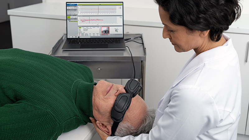
There are several steps and tools for assessing if a patient has a vestibular disorder. For a full picture of these steps, we recommend that you read this article on assessing the patient with vestibular symptoms.
A typical workflow for diagnosing patients with peripheral vestibular symptoms could look like illustrated here:

What is BPPV: Most common cause of vertigo as a result of canalithiasis (cupulolithiasis is rare).
Otoconia detach from the utricle and enter the posterior canal (~80%), the lateral canal (~18%) or the anterior canal (<2 %).
Symptoms of BPPV:
Workflow

If Dix-Hallpike identifies burst of nystagmus (typically torsional) then subsides when testing is complete, this indicates the presence of posterior canal BPPV. If Dix-Hallpike is normal (no nystagmus present), then the patient must lie on their back and turn the head to the left and then to the right. If a horizontal nystagmus in direction of the lower ear occurs (geotrophic) this is a sign of typical lateral canal BPPV. If a horizontal nystagmus in direction of the upper ear occurs (ageotrophic) this is a sign of atypical lateral canal BPPV. If no nystagmus occurs, then continue to investigate to see if the patient may have another diagnosis.
Results
Dix Hallpike: BPPV is a typical crescendo-decrescendo nystagmus observed with a delay of about 10 seconds and with a torsional component to the undermost ear and vertical upbeat component.

Impulse: Normal vHIT results confirms a diagnosis of primary BPPV. Abnormal vHIT results suggests that further tests are needed to disclose other disorders and that this is secondary BPPV.
What is Vestibular Neuritis: Acute vestibulopathy caused by inflammation of the inner ear or vestibular nerves. This inflammation disrupts the transmission of the information from the ear to the brain. This is typically viral or degenerative. Can affect the superior or the inferior branch of the vestibular nerve.
Symptoms of Vestibular Neuritis:
Workflow

If Impulse identifies catch-up saccades and cVEMP or oVEMP is abnormal then test is complete. Catch-up saccades in the lateral or anterior canals and abnormal oVEMP indicate superior vestibular neuritis. Catch-up saccades in the posterior canals and abnormal cVEMP indicate inferior vestibular neuritis.
Additional test to confirm but not necessary:

If Impulse is normal, then continue to investigate to see if the patient may have another diagnosis.
Results
Spontaneous Nystagmus: Horizontal/torsional nystagmus beating toward the good ear.


Impulse:
Pre sence of Catch-up Saccades (covert or overt) and reduced VOR gain.
cVEMP:
Reduction of amplitude on affected side.
(It should be noted that in the fields of neurology and neurophysiology convention is to have P1 (e.g. p13) as a downward deflection and N1 (i.e. n23) as an upwards deflection i.e. the reverse of what is shown below).

oVEMP:
Absent response contralateral to the lesion side while stimulating ipsilesional.
(It should be noted that in the fields of neurology and neuro-physiology convention is to have N1 (e.g. n10) as a upwards deflection i.e. the reverse of what is shown below).

Caloric:
Unilateral Weakness.

*) Ian S. Curthoys, PhD. The Interpretation of Clinical Tests of Peripheral Vestibular Function The Laryngoscope: Volume 122, Issue 6, pages 1342–1352, June 2012
Superior Vestibular Neuritis (affects the lateral and anterior canal)

Inferior Vestibular Neuritis (affects the posterior canal)


References for Benign Paroxysmal Positional Vertigo (BPPV)
Aw ST, Todd MJ, Aw GE et al (2005) Benign positional nystagmus: A study of its three-dimensional spatio-temporal characteristics. Neurology 64:1897-1905.
Baloh RW, Honrubia V, Jacobson K (1987) Benign positional Vertigo: clinical and oculographic features in 240 cases. Neurology 37:371-8.
Baloh RW, Jacobson K, Honrubia V (1993) Horizontal semicircular canal Variant of benign positional Vertigo. Neurology 43:2542-9.
Baloh RW, Yue Q, Jacobson KM et al (1995) Persistent direction? Changing positional nystagmus: another Variant of benign positional nystagmus? Neurology 45:1297-1301.
Perez-Fernandez N, Martinez-Lopez M & Manrique-Huarte R (2014) Vestibulo-ocular reflex in patients with superior semicircular canal benign paroxysmal positional Vertigo (BPPV) Acta Oto-Laryngologica. 2014; 134: 485–490.
References for Vestibular Neuritis
Akin FW, Murnane OD, Panus PC, Caruthers SK, Wilikinson AE & Proffitt TM (2004) The influence of voluntary tonic EMG level on the Vestibular evoked myogenic potential. J Rehab Res DeV 41(3B):473-480.
Aw ST, Fetter M, Cremer PD, Karlberg M, Halmagyi GM. Individual semicircular canal function in superior and inferior Vestibular neuritis. Neurology 2001;57:768–774.
Curthoys IS, Iwasaki S, Chihara Y, Ushio M, McGarvie LA & Burgess A. The ocular Vestibular-evoked myogenic potential to air-conducted sound; probable superior Vestibular nerve origin. Clinical Neurophysiology 122 (2011) 611–6.
GoVender S, Rosengren SM, Colebatch JG. Vestibular neuritis has selective effects on air- and bone-conducted cerVical and ocular Vestibular-evoked myogenic potentials. Clin Neurophysiol 2011;122:1246–1253.
Halmagyi GM, Weber KP, Curthoys IS. Vestibular function after acute Vestibular neuritis. Restor Neurol Neurosci 2010;28:37–46.
MacDougall HG, Weber KP, McGarVie LA, Halmagyi GM, Curthoys IS. The Video head impulse test: diagnostic accuracy in peripheral Vestibulopathy. Neurology 2009;73:1134–1141.
Manzari L, Tedesco AR, Burgess AM, Curthoys IS. Ocular Vestibular evoked myogenic potentials to bone conducted Vibration in superior Vestibular neuritis show utricular function. Otolaryngol Head Neck Surg 2010;143:274–280.
Manzari L, Burgess AM, MacDougall HG, Curthoys IS. Objective Verification of full recovery of dynamic Vestibular function after superior Vestibular neuritis. Laryngoscope 2011;121:2496–2500.
Manzari L, Burgess AM, Curthoys IS. Ocular and cervical Vestibular evoked myogenic potentials to bone conducted Vibration in patients with probable inferior Vestibular neuritis. J Laryngol Otol In press.
Manzari L, MacDougall HG, Burgess AM, Curthoys IS. New, fast, clinical Vestibular tests identify whether a Vertigo attack is due to early Meniere’s disease or Vestibular neuritis. Laryngoscope 2009: DOI: 10.1002/lary.23479
Pedro Luiz Mangabeira Albernaz & Francisco Carlos Zuma E Maia The Video head impulse test. Acta Oto-Laryngologica. 2014; 134: 1245–1250
Pedro L Mangabeira Albernaz & Flavia Salvaterra Cusin The Video Head Impulse Test in a Case of Suspected Bilateral Loss of Vestibular Function Int Arch Otorhinolaryngol Case Report DOI http://dx.doi.org/10.1055/s-0034-1395999
Monstad P, Okstad S, Mygland A. Inferior Vestibular neuritis: 3 cases with clinical features of acute Vestibular neuritis, normal calorics but indications of saccular failure. BMC Neurol 2006;6:45.
Shin B-S, Oh S-Y, Kim JS, et al. Cervical and ocular Vestibular-evoked myogenic potentials in acute Vestibular neuritis. Clin Neurophysiol 2012;123:369–375.
Todd NPM, Rosengren SM, Aw ST & Colebatch JG (2007) Ocular Vestibular evoked myogenic potentials (oVEMPs) produced by air- and bone-conducted sound. Clin Neurophys 118:381-390.
Weber KP, Aw ST, Todd MJ, McGarvie LA, Curthoys IS, Halmagyi GM. Head impulse test in unilateral Vestibular loss: Vestibulo-ocular reflex and catch-up saccades. Neurology 2008;70:454–463.
Weber KP, MacDougall HG, Halmagyi GM, Curthoys IS. Impulsive testing of semicircular canal function using Video-oculography. Ann NY Acad Sci 2009;1164:486–491.
Zhou G & Cox LC (2004) Vestibular evoked myogenic potentials: history and overview 13(2):135-43.