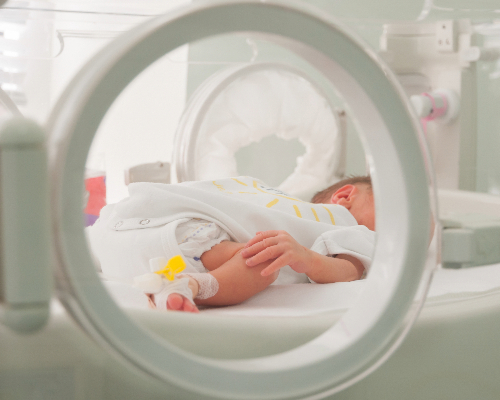
Monitoring infants has, for years, been one of the most important tasks for clinicians in the NICU. Monitoring heart rate, respiratory rate, blood pressure, temperature, etc., is standard, but yet, monitoring the brain is not a common practice.
This is despite the fact that monitoring the brain follows the same pathway, as for example, as monitoring the heart:
Another factor to consider is:
Although increases in perinatal survival rates drive clinical focus on improving long-term outcomes, many NICUs are lacking a bedside tool to help monitor neurological status. Poor neurological outcomes are associated with poor background brain activity and lack of sleep/wake cycling.
For pre-term infants, changes in aEEG patterns, such as the emergence of sleep/wake cycling, is evidence of increasing brain organization and brain health. In contrast, the absence of sleep/wake cycling or the presence of abnormal background activity is a predictor of poor long-term neurological outcomes.
In addition to background brain function, aEEG can help the clinician identify if an infant is having seizures. Eighty percent of seizures in neonates are subclinical¹, making seizure detection without aEEG monitoring difficult through clinical assessment alone. Recognizing and recording frequency and intensity of seizures is important for assisting with anticonvulsive therapy management.
aEEG helps the clinician:
aEEG was first used in the neonate population in the early 1970s for infants with hypoxic ischaemic encephalopathy (HIE). Since then, aEEG has been proven to be beneficial for additional clinical applications, such as monitoring during therapeutic hypothermia, premature infants, infants with known or suspected seizures, cardiac/surgical patients, neonatal abstinence syndrome, metabolic disease and hyperbilirubinemia.
Thus, Natus has put together a compilation of case studies to widen the perspective on the use of aEEG through the following five clinician-led cases for aEEG monitoring in neonates with the Olympic Brainz Monitor (OBM):

Related Articles