This guide for assessing patients with peripheral vestibular disorders is a quick reference of commonly diagnosed disorders. Although not a full extensive list, the guide highlights the diagnostic tools which are the most efficient and valuable for determining the presence of particular vestibular disorders.
Numerous conditions can cause a patient to experience symptoms such as dizziness or imbalance. For successful diagnosis and treatment, it is important to determine if the symptoms are due to problems in the vestibular system or a non-vestibular cause.
There are several steps and tools for assessing if a patient has a vestibular disorder, and if so, if it is a central or peripheral vestibular disorder.
Case History: This is one of the most important steps of assessing the patient. A thorough case history will assist in determining the diagnosis of the patient.
The important information is:
Note: It is necessary to rule out central causes of dizziness (e.g. stroke, traumatic brain injury, cardiovascular disease, neurological disorders (Multiple Sclerosis), anxiety, and side effects from medications or street drugs.)
Test Descriptions and Purpose: These are the common tests for assessing patients with vestibular
disorders. Which tests are performed depends on the results of the case history and the physical exam. It may also depend on your facility’s protocol.
Physical Exam (e.g. one minute eye exam): During the one minute eye exam you can have a general idea if the disorder is central or peripheral. Watch for nystagmus and pathological eye oscillations. A physician assesses the patient by asking them to watch their finger as it is moved to assess gaze, smooth pursuit and saccadic eye movement. It should also be determined if the patient has Strabismus (cover test and alternating cover test) or Internuclear opthalmoplegia.
Standard Neurological Exam: During the neurological screening you can have a general idea if the disorder is central or peripheral. This exam is an assessment of the sensory neurons and motor responses, especially reflexes, to determine whether the nervous system is impaired.
Hearing Exam: The assessment of hearing is a major step for the differential diagnosis of peripheral and central vestibulopathies and for the planning of treatment. A pure tone audiogram (with air and bone conducted stimuli) along with tympanometry and acoustic reflexes are minimally needed. The hearing assessment may also include speech testing and auditory evoked potentials. It is essential when determining if a patient has superior canal dehiscence, Menière’s disease, vestibular schwannoma or perilymph fistula.
Spontaneous/Gaze Evoked Nystagmus: The presence or absence of spontaneous nystagmus should be assessed before performing the head impulse or caloric test. Spontaneous nystagmus should be assessed without fixation by covering the eye. Recording of eye movement can be performed using the Impulse Oculomotor module or Monocular Video Frenzel functionality.
Gaze evoked nystagmus is assessed by presenting a stimulus (center and 20-30 degrees left, right, up and down) and determining if nystagmus is present. If nystagmus is present hold the gaze position for 2 minutes to determine if periodic alternating gaze is present.
For both of these tests, a video of the eye can be recorded. The video serves as documentation and can be reviewed and compared to subsequent evaluations.
VOR (VVOR & VORS): Visual VOR (vestibulo-ocular reflex) and VOR Suppression identify the presence or absence of saccadic eye movement in order to simultaneously test for the co-existence of vestibular and cerebellar pathology and thus diagnose vestibulo-cerebellar disease. VVOR assesses the patient’s vestibulo-ocular reflex with visual enhancement. VORS assesses the patient’s vestibulo-ocular reflex without visual enhancement.
Skew Deviation: Skew Deviation assesses the patient’s ocular alignment using a cover-uncover test. The purpose of this test is to identify if vertical ocular misalignment occurs as a result of covering and uncovering the eye. A vertical misalignment of the eyes is caused by a right–left imbalance of vestibular neural firing due to damage to the prenuclear vestibular input to the ocular motor nuclei affecting particularly otolithic inputs to the oculomotor system.
Saccade: Saccade assesses the visual and oculomotor system when the patient is presented with rapid goal-directed targets. The purpose of the test is to identify if abnormal eye movements are present when the patient follows the horizontal saccade stimuli. The eye movement is compared to the stimulus. Abnormal saccadic eye movement exhibited by slow, delayed saccades, result in overshoot or undershoot, lateropulsion or flutter typically indicates a central disorder.
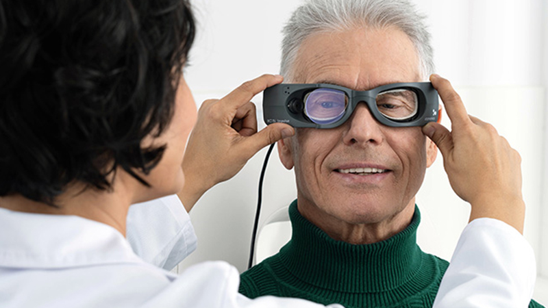
Impulse (i.e. head impulse or vHIT with SHIMP): The vHIT test is the only test that assesses all six semicircular canals independently and with a physiological stimulus, similar to how the patient uses the vestibular ocular reflex system in daily life. The test is essential in determining if the peripheral vestibulopathy affects the superior or inferior branch of the nerve, if the loss is unilateral vs bilateral, or only affecting the anterior, posterior or lateral canals. In combination with VEMPs, all 5 end organs for both ears can be assessed. Suppression Head Impulse Test (SHIMP) provides additional information regarding residual function of the vestibulo-ocular reflex system which is especially useful in patients with bilateral loss. Present catch-up saccades indicate vestibular function. No catch-up saccades present indicate vestibular loss.
Looking for a guide on how to interpret vHIT results? Download our guide to just that here.
cVEMP: Cervical Vestibular Evoked Myogenic Potential assesses the saccule using air or bone conduction stimuli. The only test used to easily assess the saccule.
oVEMP: Ocular Vestibular Evoked Myogenic Potential assesses predominantly the utricle using air or bone conduction stimuli. The completely objective test used to easily assess the utricle.
Dix-Hallpike (i.e Hallpike – Stenger) and Lateral positioning: A dynamic positional test that positions the sitting patient with their head turned 45 degrees to the left or right and then quickly moved into a supine position with the head tilted back and slightly lower than the shoulders. The purpose of the maneuver is to provoke the canaloliths to move and stimulate the canal or cupula. This is the only test that can clearly diagnose the presence of posterior canal or anterior canal BPPV (benign paroxysmal positional vertigo). Other canals must be evaluated with the head hanging and with the head lateral positions. BPPV typically exhibits a crescendo-decrescendo nystagmus observed with a delay of about 10 seconds, with a torsional component to the undermost ear and vertical upbeat component. To diagnose lateral canal BPPV, the patient must lie on
their back and turn the head to the left and then to the right.
EcochG: Electrocochleography is an electrophysiology test that assists in diagnosing cochlear hydrops by comparing the ratio of the summating potential and the action potential.
Caloric: Bithermal caloric test stimulates the left or right ear with warm and cool air or water causing a fluid density change in the lateral canal. By comparing the response of the left and right ear to warm and cool stimuli one can determine if there is a unilateral or bilateral weakness. Caloric testing is non-physiological stimulus and only assesses the lateral semicircular canal.
After assessing the patient, you may conclude that the patient’s symptoms are due to a vestibular system issue in the inner ear or vestibular nerve. However, there are several types of peripheral vestibular disorders. Learn about diagnosing some of the most common peripheral vestibular disorders.
